
The success of a root canal depends not only on how thoroughly the canals are cleaned but on how effectively the tooth is sealed afterwards. A perfectly cleaned canal system can be reinfected if bacteria from saliva and the oral environment find a way back in through an imperfect or delayed restoration. The intraorifice barrier is one of the clinical strategies used to prevent exactly that.
Understanding the Intraorifice Barrier: Definition and Purpose
The intraorifice barrier definition in clinical terms is a protective layer of material placed at the entrance of the root canals, known as the canal orifice, after the canals have been cleaned, shaped, and filled. It sits above the root canal filling material and below the final coronal restoration, functioning as a secondary seal that protects the canal system from bacterial contamination entering from above.
To understand where this layer sits, picture the internal structure of a tooth. The pulp chamber is the central cavity in the upper portion of the tooth. From there, root canals extend down through the roots. After treatment, the canals are filled from below with a material such as gutta-percha. The intraorifice barrier is then placed at the point where the canals open into the pulp chamber, sealing those openings before the permanent filling or crown is placed on top.
This is distinct from the final restoration. A crown or filling sits at the top of the tooth and restores function and appearance. The intraorifice barrier works at a deeper level, inside the pulp chamber, providing an additional line of defence that the final restoration alone does not.
Why Do Root Canal Treatments Need Extra Protection?
Coronal leakage is one of the most documented causes of root canal failure. When bacteria from the oral environment penetrate through a temporary filling, a delayed permanent restoration, or an imperfect crown margin, they can reach the canal system and recolonise it. The resulting reinfection can produce symptoms identical to the original infection and may require retreatment or, in severe cases, extraction.
A root canal intraorifice barrier reduces this risk by creating a physical seal at the canal entrance that is not dependent on the final restoration remaining intact. Even if the temporary filling is compromised between appointments, the barrier maintains a second layer of protection over the canal system.
Who Benefits Most From an Intraorifice Barrier?
| Clinical Scenario | Risk of Leakage | Barrier Commonly Considered |
| Permanent restoration delayed beyond two weeks | High | Yes |
| Temporary filling placed after treatment | Moderate to high | Yes |
| Multi-visit treatment with appointments spaced apart | Moderate | Yes |
| Immediate permanent restoration placed. | Lower | Situational |
| Patient with high caries activity | Higher | Yes |
| Single-visit treatment with same-day crown | Lower | Situational |
The decision to place a barrier is a clinical judgement made by the treating dentist based on the specific circumstances of each case. It is not a universal requirement for every root canal procedure.
Materials and Placement: How the Barrier Is Made and Positioned
Intraorifice barrier material selection is based on sealing ability, biocompatibility, handling characteristics, and the clinical situation.
| Material | Sealing Ability | Handling | Setting Time | Typical Use |
| Flowable composite | Good | Easy, syringe application | Fast, light-cured | Routine cases with tooth-coloured match |
| Glass ionomer cement | Good | Moderate | Moderate | Cases requiring fluoride release |
| Mineral trioxide aggregate (MTA) | Excellent | Requires care | Slow, moisturesensitive | Cases requiring superior biocompatibility |
| Resin-modified glass ionomer | Good | Easy | Moderate | General use |
MTA is considered the gold standard for sealing ability and biocompatibility but requires careful handling and a longer setting time. Flowable composite is widely used for its ease of application and reliable seal in routine cases. The dentist’s choice depends on the tooth, the case complexity, and the planned restoration.
How Intraorifice Barriers Are Placed
After the root canals have been cleaned, shaped, and filled with gutta-percha, the dentist prepares the canal orifice area for the barrier. Excess root filling material at the canal entrance is removed to a defined depth, creating a clean surface for the barrier material to bond to. The chosen material is applied in a controlled layer, ensuring complete coverage of each canal orifice. The seal is checked before the appointment is completed, and the barrier is then protected by the temporary or permanent coronal restoration placed on top.
The entire process adds a relatively small amount of time to the procedure but provides a disproportionate protective benefit for cases where the final restoration is delayed or where leakage risk is elevated.
Barrier, Filling and Crown: How They Work Together
The intraorifice barrier, the root canal filling, and the permanent crown or filling each serve a distinct protective function that the others cannot replicate. The root filling seals the canal system from below, preventing periapical reinfection. The intraorifice barrier seals the canal entrances from above, preventing coronal leakage. The crown or permanent filling restores function, protects the tooth structure, and provides the outermost seal.
A well-placed barrier does not reduce the importance of a timely and well-fitted permanent restoration. All three layers work together, and a weakness in any one of them creates a pathway for bacterial reinfection.
Cleaning Comes First: Irrigation and Medicaments in Root Canal Treatment
- What Is Irrigation in Root Canal Treatment?
What is irrigation in root canal treatment? It is the process of flushing the canal system with antimicrobial solutions during cleaning and shaping to remove debris, dissolve organic matter, and kill bacteria that mechanical files cannot reach.
Root canals are not simple tubes. They have lateral branches, fins, and anatomical irregularities that files cannot instrument completely. Intra canal irrigation with appropriate solutions reaches these areas through fluid dynamics, significantly improving the overall disinfection of the canal system before it is sealed.
- What Is the Best Irrigation Protocol During Root Canal Treatment?
What is the best irrigation protocol during root canal treatment is a question without a single universal answer, because the optimal approach depends on the specific tooth, the degree of infection, and the irrigant solutions available.
| Irrigant | Purpose | Key Considerations |
| Sodium hypochlorite | Dissolves organic tissue, antibacterial | Concentration and contact time matter; handle carefully. |
| EDTA | Removes the smear layer, aids penetration | Used as a final rinse before sealing |
| Chlorhexidine | Antibacterial, good substantivity | Not used with sodium hypochlorite simultaneously |
| Saline | Flushing between solutions | Does not itself disinfect |
Irrigation protocols typically involve alternating between solutions in a defined sequence, with final rinsing to remove residue before the canal is dried and filled. Activation techniques using ultrasonic or sonic devices improve the penetration of irrigants into canal irregularities. The dentist tailors the protocol to each case rather than applying a fixed sequence to every tooth.
- What Are Intracanal Medicaments?
What is intracanal medicaments in root canal treatment? They are antimicrobial substances placed inside the cleaned canal between appointments in multi-visit treatment. Their purpose is to reduce the bacterial population that remains after initial cleaning and to maintain an environment hostile to reinfection while the patient waits for the next appointment.
Calcium hydroxide is the most widely used intracanal medicament, valued for its strong antimicrobial action and its ability to dissolve residual organic tissue. It is placed as a paste or slurry, left for the prescribed period, and then completely removed before the final filling and intraorifice barrier are placed. Incomplete removal of medicament can interfere with the sealing ability of the filling material.
- What to Expect During and After the Procedure
Root canal treatment, including intraorifice barrier placement, is performed under local anaesthesia. Most patients find the procedure comfortable once anaesthesia is established. Appointment length varies depending on the number of canals and the complexity of the case.
Mild soreness for two to three days following each appointment is normal and manageable with standard over-the-counter pain relief. This reflects normal tissue healing and is not a sign of failure. Signs that warrant prompt contact with the practice include swelling, increasing pain after the third day, fever, or a persistent bad taste near the treated tooth.
Questions worth asking the dentist include which material has been used for the barrier, when the permanent restoration should be placed, and what to avoid eating during the period when only a temporary filling is in place.
Protecting Your Treated Tooth for Years to Come
A root canal-treated tooth is more brittle than a vital tooth because it no longer has a blood supply providing moisture to the dentine. Protecting it with a crown where recommended, avoiding biting hard objects such as ice or hard sweets with the treated tooth, and maintaining consistent oral hygiene reduce the risk of fracture and reinfection.
Regular dental check-ups allow the treated tooth to be monitored with X-rays over time, confirming that the periapical tissues are healing normally and that no early signs of reinfection are developing. Timely follow-up is one of the most reliable predictors of long-term success.
When Saving a Tooth Is Not Possible: Considering Replacement Options
In some cases, a tooth may be too compromised to save despite the best available treatment. Vertical root fractures, insufficient remaining tooth structure for a restoration, and advanced bone loss are among the situations in which extraction may be the recommended path.
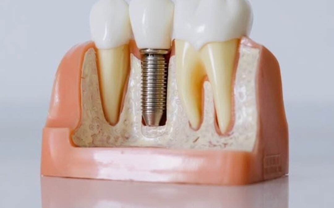
When extraction is necessary, replacement planning should begin promptly to prevent bone resorption and the shifting of adjacent teeth. Dental implants provide a permanent, stable replacement that preserves bone and restores function in a way that closely approximates a natural tooth.
Dental Magik is a trusted dental practice in East Brunswick providing experienced, personalised care across the full range of restorative and endodontic treatments. Anyone considering dental implants in East Brunswick can book a consultation with Dental Magik for a comprehensive assessment and a personalised treatment plan designed around their specific situation. Contact Dental Magik today to take the first step toward restoring your smile with confidence.
Sources:
- https://www.aae.org
- https://www.jendodon.com
- https://www.ncbi.nlm.nih.gov/pmc/articles/PMC5808543
- https://www.ese-endodontology.org
- https://onlinelibrary.wiley.com/journal/13652591
- https://www.ada.org
- https://pubmed.ncbi.nlm.nih.gov
- https://www.dental.theclinics.com
- https://www.aae.org/specialty/clinical-resources
- https://www.jcd.org.in